From chewing to chomping to grinding, teeth suffer from a lifetime of repeated mechanical stress. It makes sense, then, that enamel is one of the hardest natural materials. University of Wisconsin–Madison physics professor Pupa Gilbert and colleagues previously showed that the hydroxyapatite nanocrystals that make up enamel are arranged perfectly parallel to one another, like hairs in a ponytail, but their crystal lattices are not co-oriented — a structure that contributes to the biomaterial’s resistance to fracture, also known as toughness.
In a new study published on June 3 in the journal Nature, Gilbert and her colleagues developed a technique to quantitively measure enamel nanocrystal orientation angles across human and non-human primate enamel from different epochs, finding a strong correlation between how tough food is and the misorientation angle. The results help explain enamel evolution and have implications for modulating strength in bioinspired materials.
“Our work demonstrates that the misorientation of adjacent nanocrystals in enamel correlates very strongly with the hardness of food that primates eat,” Gilbert says. “Overall, the misorientation angles measured were small, all falling between 1.3 and 7.2 degrees, which makes sense with our earlier work where we found that small misorientation angles between thin, long, morphologically parallel nanocrystals deflect cracks and therefore toughen enamel.”
In all primates, enamel is arranged into 5-micrometer-wide bundles of elongated ~50-nanometer-wide hydroxyapatite nanocrystals. When grown synthetically, hydroxyapatite nanocrystals grow as needles, and they always have the crystalline axis along the long axis of the crystal. In enamel they do not.
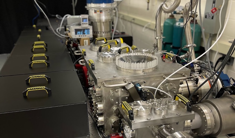
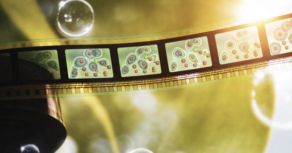
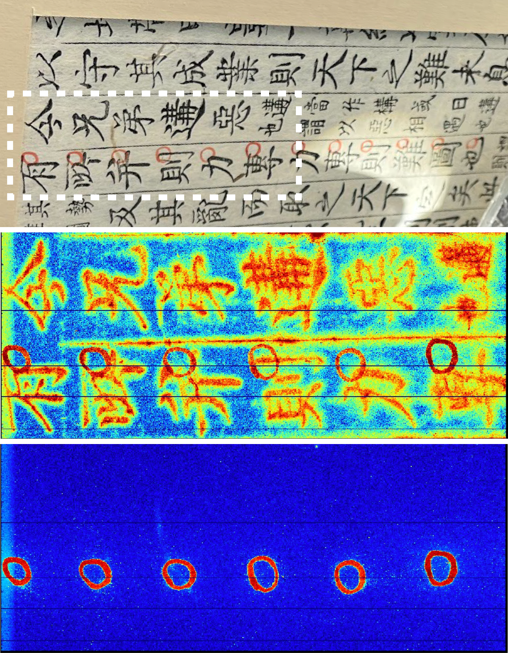
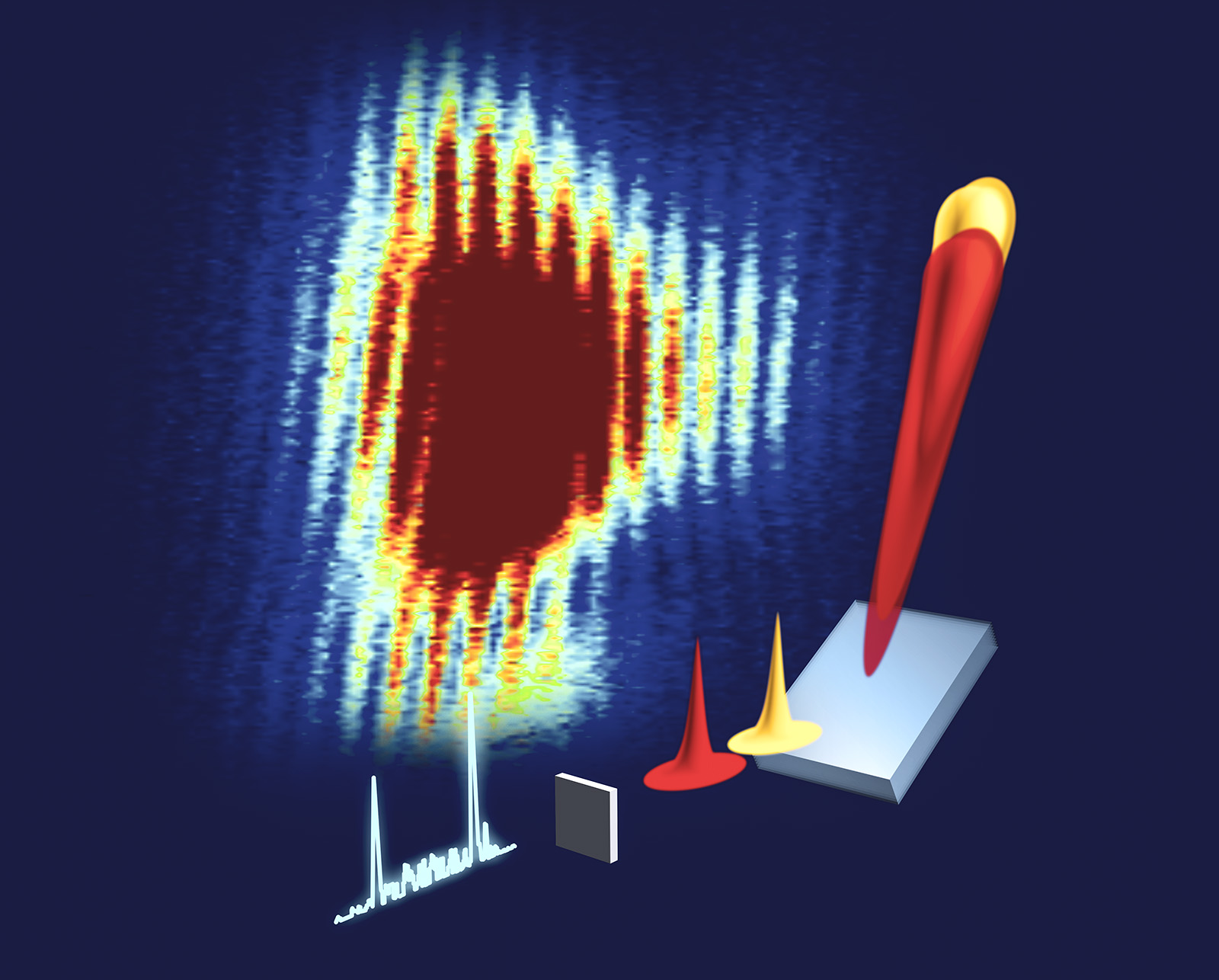
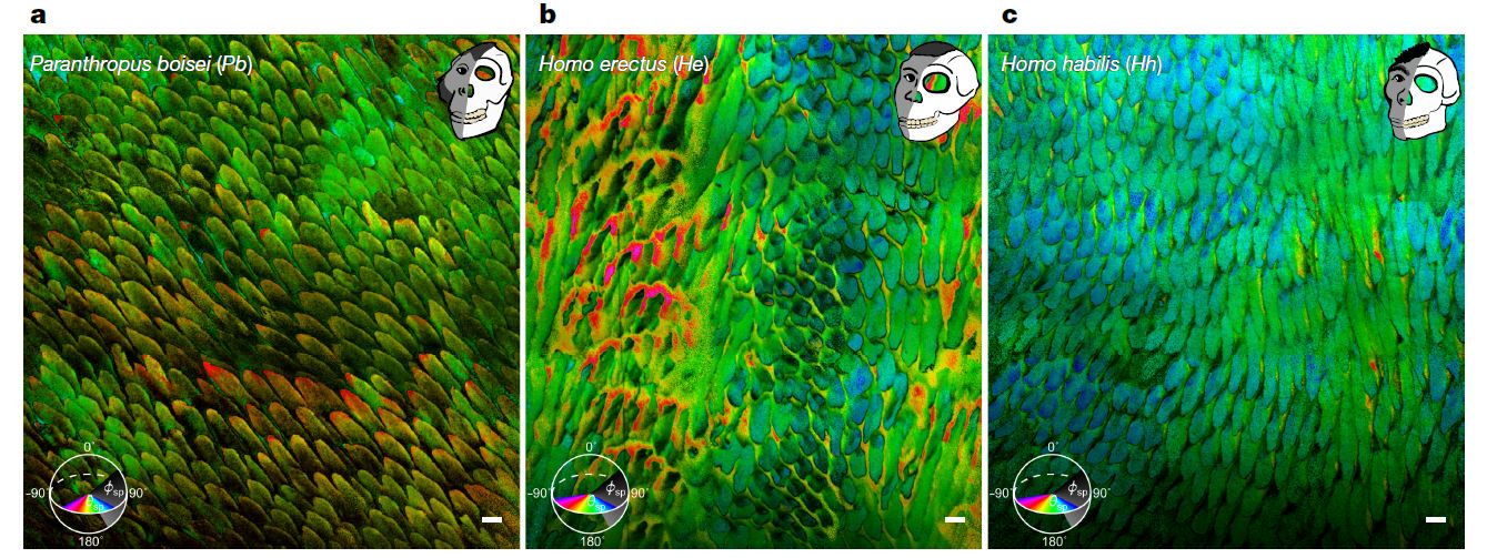
Gilbert’s new work uses a new technique she developed, called PELICAN, that displays crystal orientations quantitatively and precisely measures the misorientation of adjacent nanocrystals. This technique allows the researchers to measure the misorientation angle of one nanocrystal relative to eight neighboring crystals, with nine million angles per area. They display the data in false-colored PELICAN maps where different colors represent the range of angles, and they make histograms of the frequency of each misorientation angle.
The researchers first compared enamel structure from non-human primates, including currently living and fossilized species whose diets range from soft fruit to hard seeds and nuts. The data show a clear increase in adjacent nanocrystal misorientation angles as the primates’ food hardness increases — with a nearly six-fold increase from ripe fruit to nutshells, from 1.3 to 7.2 degrees.
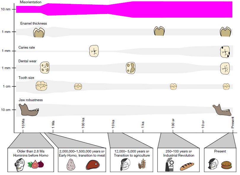
Next, they looked at primates in the human lineage, first comparing three species that lived at the same time and in the same region, ~1.6 million years ago in Kenya, but ate no meat, some meat, or mostly meat. They found that the non-meat-eater had lower misorientation angles compared to the meat eaters — 2.1 to 3-3.5 degrees — with no statistical difference between the meat eaters. Their next comparison was between Homo sapiens (paleolithic and modern humans) from before (~40,000 years ago) and after (1550 and 700 years ago) the switch to agriculture, where food in general is softer, yet they still saw an increase in crystal misorientation. However, Gilbert’s anthropologist co-author Mackie O’Hara notes that stone grinding introduced stone grit into food, making it harder and abrasive at the microscale. As in non-human primates, a general trend emerges that harder or tougher food is associated with larger misorientation angles of adjacent enamel nanocrystals.
Lastly, they looked at a modern human sample from 50 years ago, about 200 years after the Industrial Revolution when diets became much softer. Nanocrystal misorientation still went up slightly relative to the two post-agriculture Medieval samples, but the increase was not statistically significant, thus, the Industrial Revolution did not affect enamel nanostructure. Gilbert acknowledges that more research is needed to understand why misorientation angles did not decrease. One idea is that enamel adapts and evolves on a timescale greater than a few hundred years; another is that enamel is but one variable in the overall picture.
“The enamel nanostructure is only one component of a complex set of changes,” Gilbert says. “Our brains grew significantly in the last 2 million years, our jaws shrank in the last 12,000 years, we developed language, and many other changes occurred over human evolution. Even beyond genetic changes, physical characteristics change all the time, for example, crowding of the teeth toward the front of the mouth didn’t happen until after the Industrial Revolution.”
Overall, Gilbert and her team’s work suggests that primates have evolved to protect their teeth with stronger enamel as food becomes tougher. The team has not nailed down the exact misorientation angle at which maximum protection can occur, but the 1.3-7.2 degrees they measured in this study fits nicely within what materials scientists call low-angle grain boundaries, typically lower than 10-15 degrees.
“These results could also be harnessed for the synthesis of new materials that resist fracture with small misorientation of adjacent nanocrystals, such as self-assembling spherulites” Gilbert says.