by Daniel Heimsoth
Resolving very small objects that are close together is a frequent goal of scientists, making the microscope a crucial tool for research in many different fields from biology to materials science.
The resolution of even the best modern confocal microscopes — a common optical microscope popular in biology, medicine, and crystallography — is limited by an optical bound on how narrow a laser beam can be focused, known as the diffraction limit.
In a study recently published in the journal ACS Photonics, UW–Madison physics professor Shimon Kolkowitz and his group developed a method to image atomic-level defects in diamonds with super-resolution, reaching a spatial resolution fourteen times better than the diffraction limit achievable with their optics. And, because the technique uses a standard confocal microscope, this super-resolution should be available to any researchers that already have access to this common equipment.

While methods to achieve super-resolution already exist, such as stimulated emission depletion microscopy (STED), nearly all of these methods either require the addition of special optics, which can be expensive and difficult to install, or specialized samples and extensive post processing of the data. The UW–Madison technique, which they call “super-resolution Airy disk microscopy” (SAM), avoids such barriers to entry.
“You can get this all for free with the existing setup that a lot of labs already have, and it performs almost just as well,” says Aedan Gardill, a graduate student in Kolkowitz’s group and lead author of the paper. “We were able to get resolution down to twenty nanometers, which is comparable with standard techniques using [STED].”
The ‘Airy disk’ in SAM refers to a key feature of light beams that gives rise to the diffraction limit but which the researchers turned to their advantage.
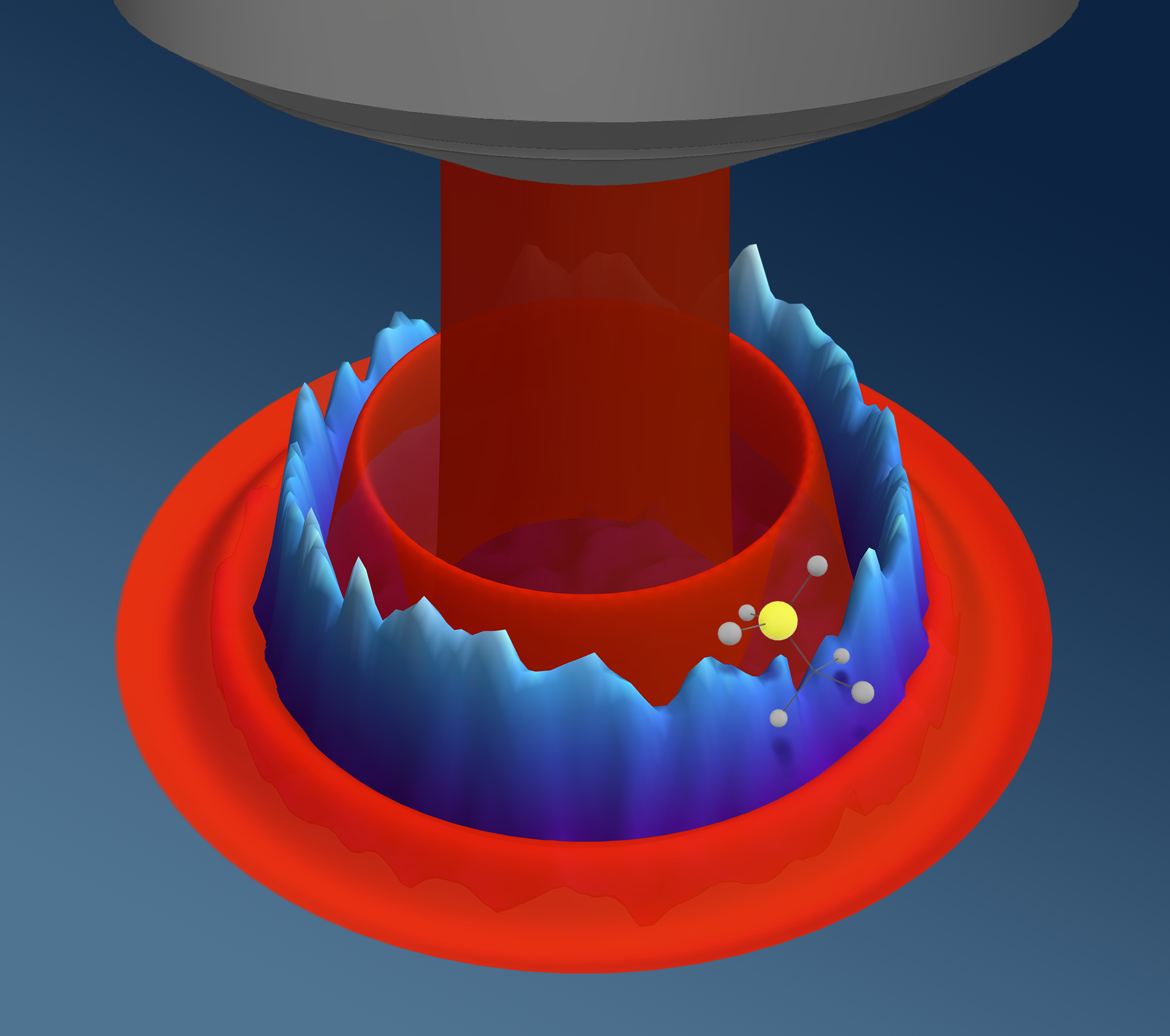
Confocal microscopes use laser beams of specific wavelengths to excite matter in a sample, causing that matter to emit light. On the microscopic scale, the laser beam does not create a solid circle of light on the sample in the same way a flashlight would. Rather, light hits the object in a series of light and dark rings called an Airy pattern. Within the dark rings, the matter receives no light, which means it cannot be detected by the microscope’s light sensors.
The novelty of the SAM technique is in its two laser beam pulses, one spatially offset from the other such that the overlapping Airy patterns can distinguish between two closely spaced objects.
In their paper, the research team studied nitrogen-vacancy (NV) centers in diamond crystal, which are regions in the crystal lattice where one of two neighboring carbon atoms is replaced by a nitrogen atom, and the other is left empty. NV centers are known to have two different charge states based on how many electrons are in the defect, one that fluoresces and one that remains dark when yellow light is applied to them.
To resolve two NV centers separated by a distance less than the diffraction limit of the microscope, the SAM procedure first shines green light on them, preparing both centers into their fluorescent charge state. Then, a red laser is applied, offset such that only one of the two NV centers is in the dark ring of the Airy pattern and thus is not affected by the beam. The NV center that does see the red light is switched to the dark state.
“It goes to another dark charge state where it does not interact with yellow light,” Gardill explains. “But the initial bright charge state does interact with yellow light and will emit light.”
Finally, when the yellow laser is applied, one NV center emits light while the other does not, effectively differentiating between the two neighboring sites. By repeating these steps iteratively over a grid, the researchers could reconstruct a full image of the two nearby NVs with spectacular resolution.
The idea for this technique came as a bit of a surprise while the team was studying charge properties of NV centers in 2020.
“We tried the combinations of red-green, green-red, red-red, green-green with those first two [laser] pulses, and the one that was green then red, we ended up seeing this ring,” Gardill recounts. “And Shimon was like, ‘The width of the ring is smaller than the size of [the confocal image of] the NV. That is super-resolution.’”
This method could find wide use in many different fields, including biology and chemistry where NV centers are used as nanoscale sensors of magnetic and electric fields and of temperature in compounds and organic material. NV centers have also been studied as candidates for quantum repeaters in quantum networks, and the research team has considered the feasibility of using the SAM technique to aid in this application. Currently, the SAM method has only been applied to NV centers in diamond crystal, and more research is needed to extend its use to different systems.
That all of this can be done with hardware that many labs across the world already have access to cannot be overstated. Gardill reiterates, “If they have a basic confocal microscope and don’t want to buy another super-resolution microscope, they can utilize this technique.”
This work was supported by the U.S. Department of Energy, Office of Science, Basic Energy Sciences under Award #DE-SC0020313.
Daniel Heimsoth is a second-year PhD student in Physics. This was his first news story for the department.